Injuries to the meniscus
A full range of orthopedic services, from diagnosis to full recovery
Menisci are semilunar cartilaginous formations located inside the knee joint.
Right knee joint, front view, without patella.
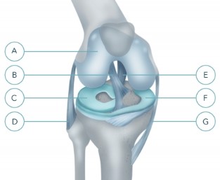
A - articular cartilage
B - anterior cruciate ligament
C - external (lateral) meniscus
D - peroneal (external) collateral ligament
E - posterior cruciate ligament
F - internal (medial) meniscus
G - tibial (internal) collateral ligament
The human knee joint is a complex biomechanical structure. It is formed by the articular surfaces of the femur, tibia, and patella. Inside the joint, the surfaces of the bones are covered with cartilage.
All the elements are held together by the joint capsule and ligaments.
Between the articular surfaces of the tibia and femur, there are cartilaginous formations – external and internal menisci. The inner surface of the joint capsule is covered with a mucous membrane. The ligaments are located inside and outside the joint: inside there are two cruciate ligaments (anterior and posterior), outside-the collateral ligaments.
Question: What functions do the different structures of the joint perform?
Cartilage is the tissue that covers the intra-articular surfaces of the bones and provides free, painless movement in the knee joint. The cartilage does not contain blood vessels. Its nutrition comes from the synovial fluid that fills the joint cavity. Synovial fluid is produced by the mucous membrane of the joint capsule.
Menisci:
Q: What is the cause of meniscus tears?
There are two reasons:
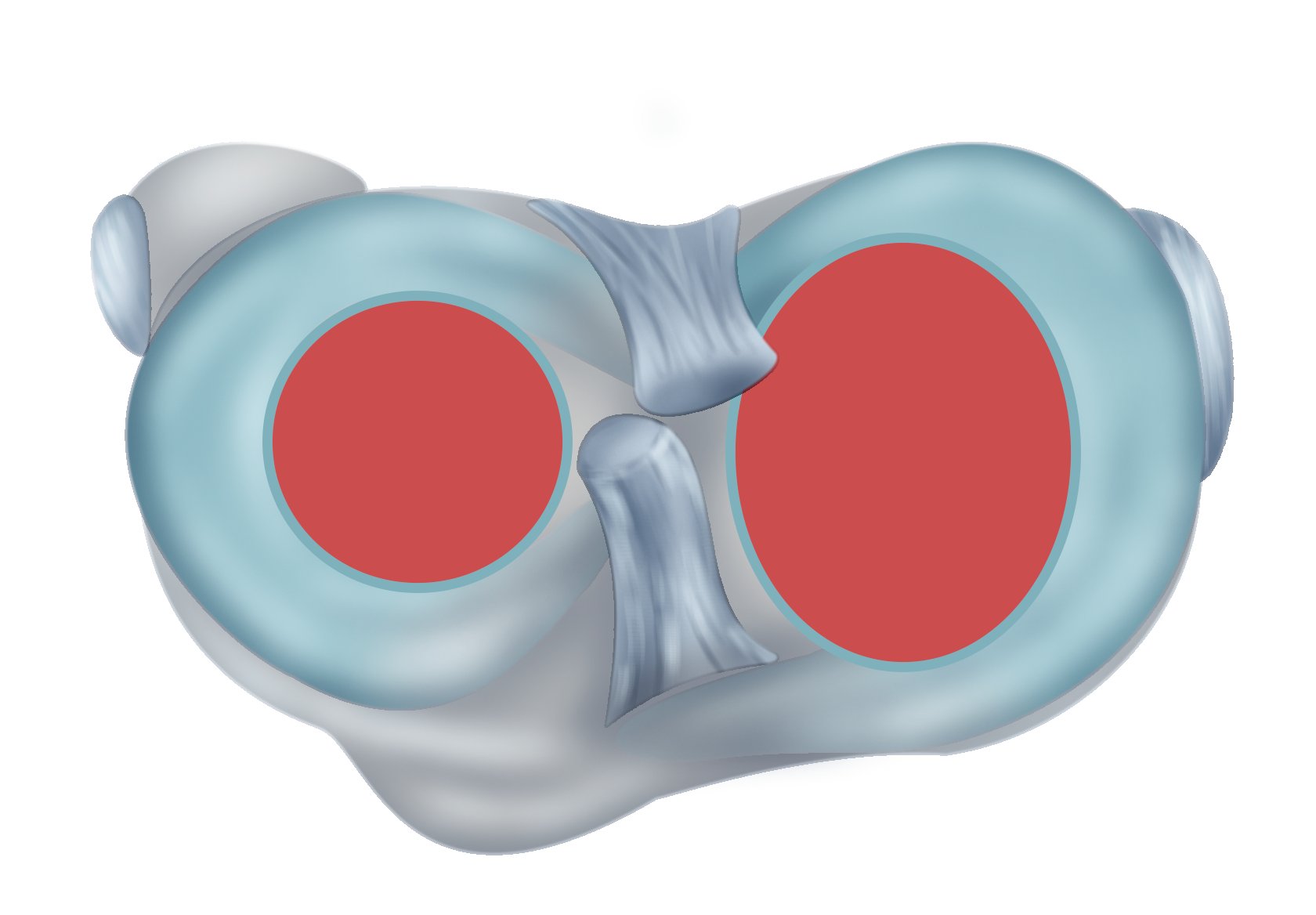
Fig. 2. The area of contact between the bones of the knee joint on the articular surface of the right tibia - indicated in red (top view).
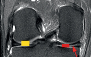
3. Horizontal rupture (indicated by an arrow) of the internal meniscus due to compression of the bones as a result of their convergence against the background of a decrease in the height of the articular cartilage (indicated by a red square).
The distance between the bones depends on the total thickness of the articular cartilage that these bones cover. The bones have different shapes, and there is a space between them, which is occupied by the menisci (Fig. 3). Due to overload, the total thickness of the cartilage decreases, the bones begin to converge. There is compression of the menisci, causing necrosis of their central part, followed by cracking and the formation of a rupture line (the so-called "degenerative rupture"). Both menisci in one person are rarely damaged. The rupture usually occurs from the side of a greater decrease in the thickness of the cartilage.
Degenerative ruptures can occur spontaneously, when performing normal daily activities.
Q: What are the symptoms of a meniscus tear?
Typical:
Damage to the ligaments and menisci dramatically disrupts the normal biomechanics of the joint, which leads to an uneven load on the articular cartilage. Those parts of the cartilage that experience an increased load begin to quickly collapse (Fig. 2). Damage to the articular cartilage is an irreversible process that leads to the development of deforming arthrosis and impaired function of the knee joint as a means of transportation.
Q: How can I confirm the diagnosis of meniscus injury?
The diagnosis is based on:
Patients with meniscal rupture in the vast majority need surgery (the exception is the presence of contraindications to surgical interventions, old age).
Question: what is the essence of the operation?
In the medical center "Orthopedics of Ruslan Sergienko" all operations for traumatic injuries and diseases of the knee joint are performed arthroscopically-through several punctures of the skin above the joint, under the control of a video camera.
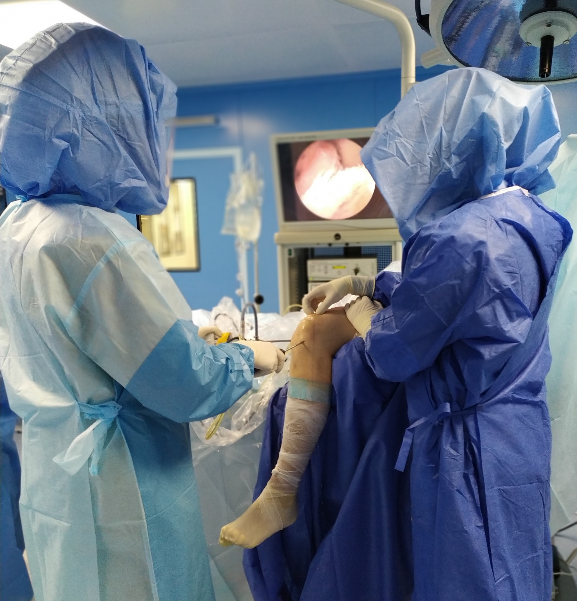
Arthroscopic Knee surgery: The surgeon performs the operation inside the joint while looking at the monitor.
Question: Why is arthroscopic surgery better than open surgery?
Modern intraoperative monitoring minimizes the likelihood of" waking up " during general anesthesia.
Question: what is the essence of the operation for the meniscus injury?
Operations are divided into 2 types:
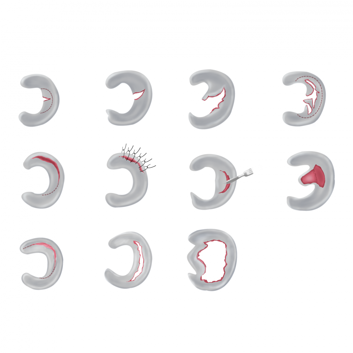
The meniscus is an important part of the joint, and it is important to preserve it at the least possible opportunity.
The blood supply to the meniscus is most developed in its peripheral parts, located along the edge of the joint, near the joint capsule. These same parts of the meniscus are most functionally significant.
Right knee joint, front view, without patella.

A - articular cartilage
B - anterior cruciate ligament
C - external (lateral) meniscus
D - peroneal (external) collateral ligament
E - posterior cruciate ligament
F - internal (medial) meniscus
G - tibial (internal) collateral ligament
The human knee joint is a complex biomechanical structure. It is formed by the articular surfaces of the femur, tibia, and patella. Inside the joint, the surfaces of the bones are covered with cartilage.
All the elements are held together by the joint capsule and ligaments.
Between the articular surfaces of the tibia and femur, there are cartilaginous formations – external and internal menisci. The inner surface of the joint capsule is covered with a mucous membrane. The ligaments are located inside and outside the joint: inside there are two cruciate ligaments (anterior and posterior), outside-the collateral ligaments.
Question: What functions do the different structures of the joint perform?
Cartilage is the tissue that covers the intra-articular surfaces of the bones and provides free, painless movement in the knee joint. The cartilage does not contain blood vessels. Its nutrition comes from the synovial fluid that fills the joint cavity. Synovial fluid is produced by the mucous membrane of the joint capsule.
Menisci:
- increase the correspondence of the articular surfaces of the tibia and femur;
- increase the contact area between them;
- reduce the load on the cartilage of the articular surfaces of the bones;
- dampen movement under vertical loads-running, jumping;
- together with the ligaments create mechanical strength and stability of the joint;
- provide a uniform load on the articular cartilage, maintaining its integrity throughout life.
Q: What is the cause of meniscus tears?
There are two reasons:
- knee injury - during uncontrolled flexion or twisting, often combined with ligament damage;
- the consequences of overloading - a more complex mechanism: the bones of the knee joint contact each other on a small area of the articular surface (Fig. 2). The rest of the contact occurs through the surface of the meniscus.

Fig. 2. The area of contact between the bones of the knee joint on the articular surface of the right tibia - indicated in red (top view).

3. Horizontal rupture (indicated by an arrow) of the internal meniscus due to compression of the bones as a result of their convergence against the background of a decrease in the height of the articular cartilage (indicated by a red square).
The distance between the bones depends on the total thickness of the articular cartilage that these bones cover. The bones have different shapes, and there is a space between them, which is occupied by the menisci (Fig. 3). Due to overload, the total thickness of the cartilage decreases, the bones begin to converge. There is compression of the menisci, causing necrosis of their central part, followed by cracking and the formation of a rupture line (the so-called "degenerative rupture"). Both menisci in one person are rarely damaged. The rupture usually occurs from the side of a greater decrease in the thickness of the cartilage.
Degenerative ruptures can occur spontaneously, when performing normal daily activities.
Q: What are the symptoms of a meniscus tear?
Typical:
- pain on the inner or outer surface of the knee, especially pronounced in the squat;
- "flipping";
- it is often impossible full flexion and extension;
- sudden blocking of movements in the knee;
- swelling of the knee.
Damage to the ligaments and menisci dramatically disrupts the normal biomechanics of the joint, which leads to an uneven load on the articular cartilage. Those parts of the cartilage that experience an increased load begin to quickly collapse (Fig. 2). Damage to the articular cartilage is an irreversible process that leads to the development of deforming arthrosis and impaired function of the knee joint as a means of transportation.
Q: How can I confirm the diagnosis of meniscus injury?
The diagnosis is based on:
- clinical study (study of complaints, mechanism of injury, examination of the knee joint with the help of special tests);
- magnetic resonance imaging (required).
Patients with meniscal rupture in the vast majority need surgery (the exception is the presence of contraindications to surgical interventions, old age).
Question: what is the essence of the operation?
In the medical center "Orthopedics of Ruslan Sergienko" all operations for traumatic injuries and diseases of the knee joint are performed arthroscopically-through several punctures of the skin above the joint, under the control of a video camera.

Arthroscopic Knee surgery: The surgeon performs the operation inside the joint while looking at the monitor.
Question: Why is arthroscopic surgery better than open surgery?
- The image on the monitor is enlarged, which allows you to perform surgery more accurately.
- Tools for arthroscopic operations of small sizes, which improves the quality of the operation and reduces injury to the joint tissues.
- The operation takes place without contact of the joint with the environment, which reduces the risk of postoperative infection.
- Without injuring the soft tissues with surgical hooks to dilute the edges of the wound.
- Accelerated healing period and length of hospital stay.
- Post-operative scarring after healing is much less noticeable – women will be able to wear short skirts without stress.
- The patient is assigned to the date of hospitalization;
- prior to surgery, the patient undergoes tests and makes the survey a complete list of which is determined individually;
- hospitalization on the morning of the operation;
- examination by a physician anesthesiologist, the decision about the optimal method of anesthesia;
- the patient is taken to the operating unit, the anesthesiologist performs intravenous administration of drugs for anesthesia or spinal anesthesia.
- The use of anesthesia in our time is not dangerous for the patient's body.
- During anesthesia, the doctor constantly monitors the vital signs and the amount of drugs, depending on the duration of the surgical intervention.
- It is impossible to wake up during the operation.
- After the operation, after a certain time, the effect of the drug ends, and the patient wakes up.
- During spinal anesthesia, a local anesthetic solution is injected into the subarachnoid space located in the spinal canal. As a result, there is anesthesia of the lower half of the body or one leg.The introduction of a local anesthetic is carried out below the end of the spinal cord, the injury of the latter during spinal anesthesia is excluded. The dose of the anesthetic is so small that it can be compared to anesthesia in dentistry. 3-7 minutes after the introduction of a local anesthetic, there is a temporary blockage of the nerve fibers that innervate the lower half of the body or one leg, which is accompanied by a feeling of warmth, numbness, decreased sensitivity and relaxation of the muscles of the lower extremities. The duration of action, depending on the chosen anesthetic, can be from 4 to 6 hours.
Modern intraoperative monitoring minimizes the likelihood of" waking up " during general anesthesia.
Question: what is the essence of the operation for the meniscus injury?
Operations are divided into 2 types:
- removal (resection) of the damaged part of the meniscus;
- suturing of the damaged part of the meniscus.
- the type of rupture,
- the presence of blood circulation at the site of the rupture,
- the age of the injury.

The meniscus is an important part of the joint, and it is important to preserve it at the least possible opportunity.
The blood supply to the meniscus is most developed in its peripheral parts, located along the edge of the joint, near the joint capsule. These same parts of the meniscus are most functionally significant.
Product rating is: 0 from 5
Please rate the product:
Advantages of treatment in Orthopedics by Ruslan Sergienko
-
10 years on medical services in Ukraine
-
> 25 years of experience with leading specialists
-
Anna Vovchenko and Ruslan Sergienko are recognized opinion leaders among the orthopedic traumatologists
-
> 150,000 consultations were held
-
> 7,500 surgeries were performed
-
All types of pain management
-
The operating unit is equipped according to international standards
-
Availability of all medicines and supplies
-
Single rooms, equipped with the characteristics of orthopedic patients
-
Three meals a day
-
Postoperative rehabilitation by certified specialists
-
Pricing Transparency
