Menu

Ultrasound examination of the musculoskeletal system

Ultrasound

modern, informative method of joint research using ultrasound waves.

In Ukraine, ultrasound examination of the musculoskeletal system has been used since the late 90s of the last century, and during this time it has become firmly established in medical practice and has become an integral part of diagnostics in traumatology and orthopedics, sports medicine and rheumatology.

Advantages of ultrasound examination of joints:

unlike X-ray examination, which allows you to see only bone structures, ultrasound examination of joints is aimed at visualizing the soft tissues of the joints (muscles, ligaments, cartilage, tendons,
capsules), invisible on a normal X-ray image. 


Ultrasound examination of joints and soft tissues:


  • Ultrasound examination of the joints is an absolutely safe method, it does not contain harmful ionizing radiation, which is why ultrasound can be done an unlimited number of times.
  • Ultrasound examination of the joints in many research issues successfully competes with MRI and CT, but it is a more affordable method, its cost is several times less than that of MRI and CT studies.
  • Ultrasound examination allows you to evaluate changes in the joints in dynamics (for example, the state before and after treatment).
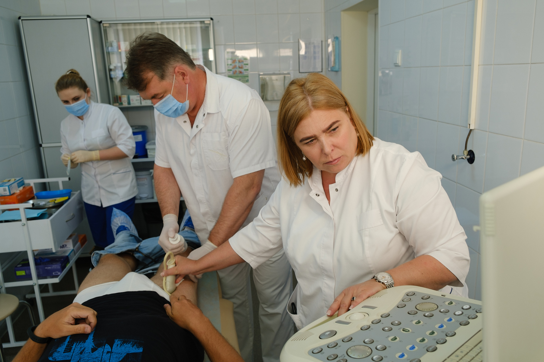
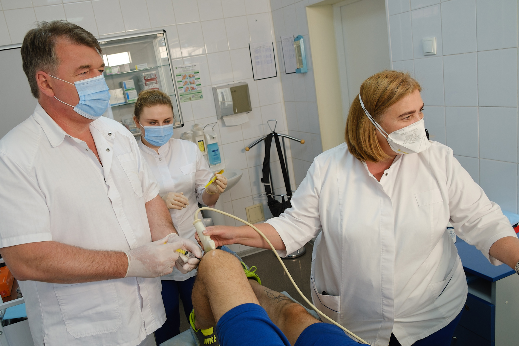
  • Ultrasound examination allows for visual control of the introduction of drugs into the joint cavity and periarticular tissues (so-called ultrasound navigation).

Who performs joint ultrasound in our medical center?


In our medical center, ultrasound examination of the joints is carried out Anna Yakovlevna Vovchenko is one of the leading specialists of Ukraine in the field of ultrasound diagnostics of the musculoskeletal system. One of the first in Ukraine to introduce the method of ultrasound examination of joints into clinical practice. In 1993, she was trained in Austria by the world-famous Professor R. Graf, and since then she has taught this method to more than 200 doctors in Ukraine. He has extensive practical experience in the ultrasound diagnosis of joint diseases, is the author of books and monographs on ultrasound of the musculoskeletal system.

Realities and prospects of orthopedic sonography presentation by Anna Yakovlevna Vovchenko

Features of joint ultrasound in our medical center:


In our medical center, ultrasound examination of all joints is performed: shoulder, elbow, hip, knee, small joints of the hands and feet, as well as ultrasound of the hip joints in children of the first year of life, if hip dysplasia is suspected.

Ultrasound examination necessarily includes an examination of two symmetrical joints (two knee, two elbow, etc.), since a comparative assessment of the structures of the affected joint with the opposite "healthy" joint is necessary. Examination of symmetrical joints also allows you to diagnose pathological changes in a "healthy" joint, which does not yet bother the patient, but it is already in the initial development of pathology.

We use ultrasound examination during punctures, intra-articular and periarticular injections - to control the correct entry into the joint and the administration of the optimal dose of the drug. Under the control of ultrasound, needling is also performed – a procedure aimed at washing out calcinate in the tendons. In some cases, ultrasound is performed during arthroscopic operations to better visualize the pathology (for example, calcinate in muscle tendons).

Intra-articular injections under ultrasound control


DSCF9994.jpg
DSCF0171.jpg

DSCF9831.jpg

Patients ' questions about joint ultrasound:

Why do an ultrasound if an x-ray is taken?
Traditional radiographic examination allows to evaluate only the bone structures of the joint, but does not visualize the soft tissues of the joint and the inflammatory fluid.

Why perform an ultrasound of two symmetrical joints if one hurts?
Ultrasound examination necessarily includes an examination of two symmetrical joints (two knee, two elbow, etc.), since a comparative assessment of the structures of the affected joint with the opposite "healthy" side is necessary. When examining symmetrical joints, very often pathological changes are found in the "healthy" joint, although there may be no clinical symptoms: pain, discomfort or poor mobility.

Is it harmful to perform ultrasound for infants and pregnant women?
Ultrasound is an absolutely safe diagnostic method that can be safely prescribed to young children and women at any stage of pregnancy.

If the knee (shoulder, elbow, or any other joint) hurts, but there was no injury, then which diagnostic method is better to start the examination: X-ray, ultrasound, or MRI?
If there is pain in the joints, and there was no injury, it is better to start with an ultrasound. More than 80% of joint pain that is not associated with trauma is inflammatory or degenerative-dystrophic diseases of the joints. In this case, ultrasound can show changes in the articular cartilage, the presence of signs of inflammation, loose bodies in the joint cavity, and much more.

Advantages of treatment in Orthopedics by Ruslan Sergienko

  • 10 years on medical services in Ukraine
  • > 25 years of experience with leading specialists
  • Anna Vovchenko and Ruslan Sergienko are recognized opinion leaders among the orthopedic traumatologists
  • > 150,000 consultations were held
  • > 7,500 surgeries were performed
  • All types of pain management
  • The operating unit is equipped according to international standards
  • Availability of all medicines and supplies
  • Single rooms, equipped with the characteristics of orthopedic patients
  • Three meals a day
  • Postoperative rehabilitation by certified specialists
  • Pricing Transparency

Feed back

This website uses cookies to make your use of the site more user-friendly.
Learn more about cookies and cookie policies. By clicking OK, you agree with it.